
Page 48 -
P. 48
ิ
์
ิ
ิ
ิ
ื
ุ
ั
โครงการพัฒนาหนังสออเล็กทรอนกสเฉลมพระเกียรต สมเด็จพระเทพรตนราชสดาฯ สยามบรมราชกุมาร ี
45
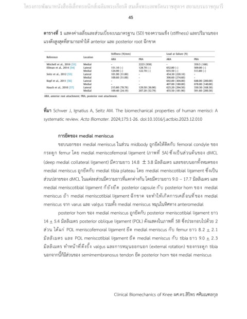
ตารางที่ 1 แสดงค่าเฉลี่ยและส่วนเบี่ยงเบนมาตรฐาน (SD) ของความแข็ง (stiffness) และปริมาณของ
แรงดึงสูงสุดที่สามารถทำให้ anterior และ posterior root ฉีกขาด
ที่มา Schwer J, Ignatius A, Seitz AM. The biomechanical properties of human menisci: A
systematic review. Acta Biomater. 2024;175:1-26. doi:10.1016/j.actbio.2023.12.010
การยึดของ medial meniscus
ขอบนอกของ medial meniscus ในส่วน midbody ถูกยึดให้ติดกับ femoral condyle ของ
กระดูก femur โดย medial meniscofemoral ligament (ภาพที่ 5A) ซึ่งเป็นส่วนต้นของ dMCL
(deep medial collateral ligament) มีความยาว 14.8 3.8 มิลลิเมตร และขอบนอกทั้งหมดของ
medial meniscus ถูกยึดกับ medial tibia plateau โดย medial meniscotibial ligament ซึ่งเป็น
ส่วนปลายของ dMCL ในแต่ละส่วนมีความยาวที่แตกต่างกัน โดยมีความยาว 9.0 – 17.7 มิลลิเมตร และ
medial meniscotibial ligament ก็ยังยึด posterior capsule กับ posterior horn ของ medial
meniscus ถ้า medial meniscotibial ligament ฉีกขาด จะทำให้เกิดการเคลื่อนที่ของ medial
meniscus จาก varus และ valgus รวมทั้ง medial meniscus หมุนในทิศทาง anteromedial
posterior horn ของ medial meniscus ถูกยึดกับ posterior meniscotibial ligament ยาว
14 + 5.4 มิลลิเมตร posterior oblique ligament (POL) ดังแสดงในภาพที่ 5B ซึ่งประกอบไปด้วย 2
ส่วน ได้แก่ POL meniscofemoral ligament ยึด medial meniscus กับ femur ยาว 8.2 + 2.1
มิลลิเมตร และ POL meniscotibial ligament ยึด medial meniscus กับ tibia ยาว 9.0 + 2.3
มิลลิเมตร ทำหน้าที่ดึงรั้ง valgus และการหมุนออกนอก (external rotation) ของกระดูก tibia
นอกจากนี้ก็มีส่วนของ semimembranosus tendon ยึด posterior horn ของ medial meniscus
Clinical Biomechanics of Knee ผศ.ดร.สิริพร ศศิมณฑลกุล